How do doctors diagnose emphysema? - Medical News Today

Doctors can diagnose emphysema using several tests, including spirometry. This test measures how much air a person can exhale from their lungs after taking a deep breath. Other tests may include imaging scans and blood tests.
Emphysema is a severe respiratory disease that damages the air sacs, or alveoli, of the lungs. It can also affect the walls between them. This damage causes airways to become inflamed and rigid, which leads to difficulty breathing and other serious health concerns.
Emphysema falls under the umbrella of chronic obstructive pulmonary disease (COPD), along with chronic bronchitis.
Continue reading to learn more about how doctors diagnose emphysema.
Doctors often diagnose emphysema without using routine laboratory tests or imaging studies. Instead, they use lung function tests as the
Diagnosis typically begins by taking a detailed medical history to assess an individual's current symptoms and any previous or current medical conditions. This allows the doctor to determine any potential underlying causes or risk factors for emphysema.
Next, the doctor will perform a physical exam to check how well the lungs and heart are functioning. This includes:
- listening to the lungs with a stethoscope
- feeling for specific areas of tenderness or swelling
- checking whether there is an accumulation of fluid in the lungs
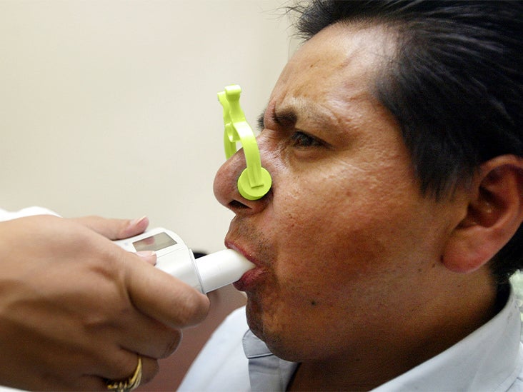
Finally, the doctor may perform pulmonary function tests. This may include spirometry, which is the most common test that doctors use to diagnose COPD. It measures how much air a person can exhale after taking a deep breath and how quickly they exhale.
The results of spirometry will help the doctor:
- determine how severe the COPD is
- check how well the lungs are working
- make treatment recommendations
To diagnose emphysema, a doctor may perform body plethysmography and a diffusing capacity test. Body plethysmography measures the amount of air in the lungs, and a diffusing capacity test measures how efficiently oxygen passes from capillaries in the lungs to hemoglobin in the bloodstream.
Taking a thorough medical history helps the doctor understand how long a person has been experiencing symptoms and how those symptoms have evolved over time. This can help rule out other conditions that may have similar symptoms, such as asthma or chronic bronchitis.
The doctor will ask questions about:
- the type and duration of cough
- whether there is lots of phlegm or mucus
- whether specific activities cause breathlessness
- how the symptoms impact daily life
- whether the person's family has a history of lung disease
- whether they have a personal history of:
- chest problems
- allergies
- infections
- smoking
- exposure to irritants or toxins, such as dust, fumes, or chemicals
- other medical conditions
Doctors also ask about any regular medications the individual is taking. Some medications may affect lung capacity and functioning.
The next step of diagnosis is a physical examination. This can help a doctor check how well the lungs are functioning and the degree of any obstructions. It can also help detect signs of other conditions that could be causing the symptoms.
During a physical exam, the doctor listens to the lungs using a stethoscope, which is called auscultation. This can help them determine how much air is flowing through the lungs, how fast it is moving, and whether there are areas of decreased breath sounds due to blockages caused by inflammation.
In addition, doctors can use this test to listen for any atypical sounds, such as wheezing or crackling, which would indicate airway issues.
Next, a doctor may try percussion, or tapping on the chest. In combination with auscultation, this may yield evidence of left heart failure or pleural effusion, which is an accumulation of fluid in the lungs.
Testing vital signs, including pulse rate and oxygen saturation levels, may allow a doctor to determine how well the body can maintain oxygen levels.
Imaging tests such as CT scans and MRI scans are not part of the routine evaluation for suspected COPD. In some cases, doctors may use chest X-rays to help them with their diagnosis and rule out any other potential conditions.
However, typically a chest X-ray is
X-rays may also help rule out other conditions.
Laboratory tests, such as blood tests, cannot confirm emphysema or COPD. However, doctors may order tests in some cases.
For example, if a young person has symptoms of emphysema, they
Doctors may also order arterial blood gas analysis to measure how much oxygen and carbon dioxide are present in the body. This informs the doctor how severe the condition is and how well the lungs are functioning.
Lung function tests are a primary tool for COPD diagnosis. Spirometry is the most common test that doctors use for diagnosis. It reveals how much air an individual can breathe out in one breath, how deep a breath they can take, and how quickly this happens.
The results of spirometry will help the doctor determine how severe the COPD is and check how well the lungs are working.
Spirometry involves inhaling deeply, then exhaling as much air as possible into a device that measures how quickly and how much air is expelled from the lungs.
The doctor predicts typical values for the individual based on their age, sex, and height. They then compare test scores to this figure.
The device records the results in measurements known as forced vital capacity (FVC) and forced expiratory volume in one second (FEV1). The doctor looks at these measurements individually and as a combined value known as the FEV1/FVC ratio.
If the FEV1/FVC ratio is less than 70%, this suggests that COPD is present. An FEV1 greater or equal to
Another lung function test doctors may use is a diffusing capacity test. For this, a person wears a mouthpiece and inhales a small amount of gas. Doctors can then measure how quickly the gas moves from the lungs to the bloodstream.
Diagnosing emphysema involves taking a detailed medical history, performing a physical exam, and listening to the lungs.
Pulmonary function testing is the main diagnostic tool for checking how severe a person's emphysema is and how well their lungs are functioning.
In some cases, doctors may order imaging tests or laboratory tests to check for other potential causes. However, an X-ray cannot confirm COPD. Only spirometry can do this.
Getting an accurate diagnosis is important because it guides the treatment and management of symptoms.

Comments
Post a Comment